
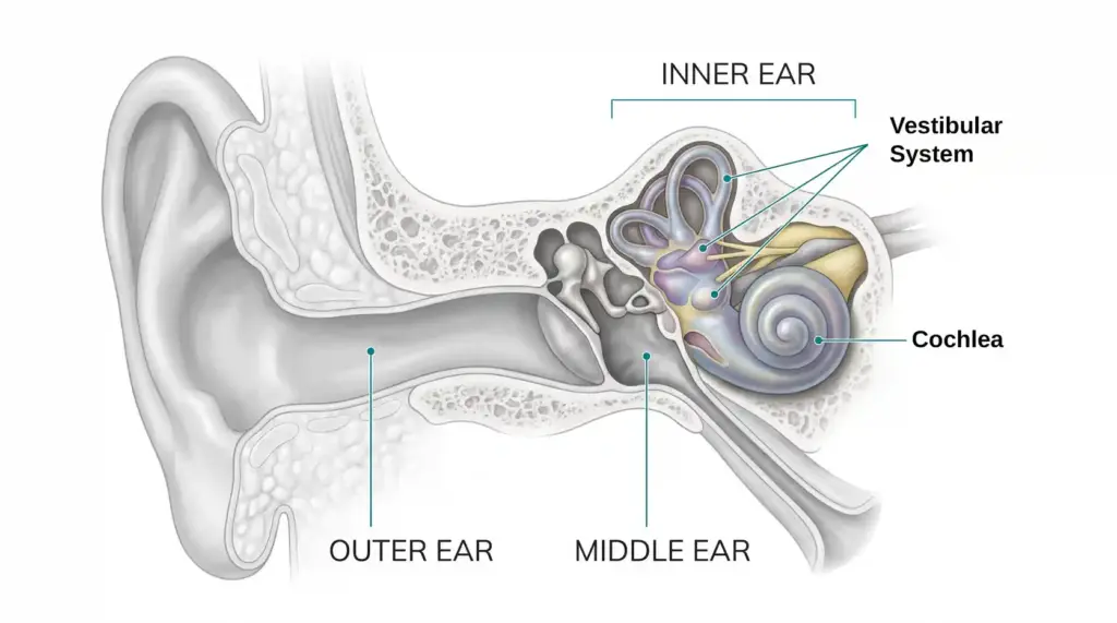
The inner ear is the innermost part of the ear, containing structures responsible for hearing, balance, and spatial orientation.
It includes the cochlea, which is responsible for hearing, and the vestibular system, which contributes to balance, spatial orientation, and stabilizing gaze during head movement. These structures are housed within the temporal bone of the skull.
Within the cochlea, specialized sensory cells convert mechanical vibrations into electrical signals. These signals travel through the auditory nerve to the brain, where they are interpreted as sound. Signals from the vestibular system travel along vestibular nerve pathways to help the brain maintain balance and coordinate movement.
The inner ear can be stimulated through both air conduction and bone conduction. In air conduction, vibrations reach the cochlea through the usual pathway involving the outer ear, eardrum, and ossicles. In bone conduction, vibrations transmitted through the skull stimulate the cochlea without relying on this usual pathway.
QUICK FACT
The cochlea and auditory nerve are essential for normal hearing through both air and bone conduction. Bone conduction can therefore help assess inner-ear hearing function when a problem with the outer or middle ear is suspected.
COMMONLY CONFUSED WITH
- Middle ear: Transmits sound vibrations through the eardrum and ossicles, whereas the inner ear contains the sensory structures that convert mechanical vibrations into neural signals.
- Bone conduction: A way of transmitting sound vibrations to the cochlea through the bones of the skull rather than relying on the usual air-conduction pathway.
SEE ALSO